Dr. Alberto Macciò
Specialist in General Surgery
Perfected in Microsurgery and Lymphology

Dr. Alberto Macciò
Specialist in General Surgery
Perfected in Microsurgery and Lymphology

In our center, by virtue of decades of experience in the field of lymphatic circulation diseases, we mainly deal with complex diagnoses and complications.
Very often patients do not achieve full therapeutic success because of the lack of an accurate and circumstantial diagnosis. In our Center, by making use of the most modern diagnostic methods for lymphological patient framing (Lymphoscintigraphy, 3d Perometry, Lymphography, LymphangioRM, Lymphoscintigraphy with Spet CT, genetic testing) we are able to best meet therapeutic needs.
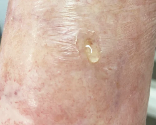
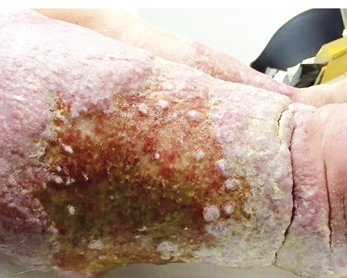
Given our surgical training, most of our daily activity has now focused for more than a decade on the treatment of acute complications of the lymphedema patient such as:
Diagnostic methods for the framing of the lymphological patient
Lymphoscintigraphic examination *
The gold standard of lymphatic circle assessment is segmental limb lymphoscintigraphy. By distally injecting albumin nanocolloids labeled with Tc-99m (radioisotope), functional lymphatic drainage pathways can be verified in detail and dynamically. A relatively minimally invasive examination and allows excellent imaging of even the satellite lymphatic-lymph node districts, highlighting any areas of stop or dermal back flow (superficial dermal reflux typical of advanced stages of pathological lymph stasis). Segmental limb lymphoscintigraphy is performed in the Hospital, in the Department of Nuclear Medicine.
* Bourgeois P., Munck D., Belgrade J.P., Leduc O., Leduc A.(2003) Limb edema and Lymphoscintigraphy.Rev Med Brux Feb;24(1):20-8.
LymphangioRMN
It consists of imaging the lymphatic system after administration of Gadolinium. It allows a more refined analysis of the anatomical condition of the loco-regional lymphatic system. LymphangioRMN is performed in special diagnostic centers.
3D Perometry *
The Bodytronic® 600 system provides rapid, highly reliable and accurate measurements of lower limb circumference and volume. It can be considered a valuable measurement tool for use in various clinical and research applications. 3D Perometry analysis is performed with the system the Bodytronic® 600 system at our Studio at each visit. (VIDEO)


